You are here
lymphocyte-attacking-cancer-cell.jpg

Media Folder
Title
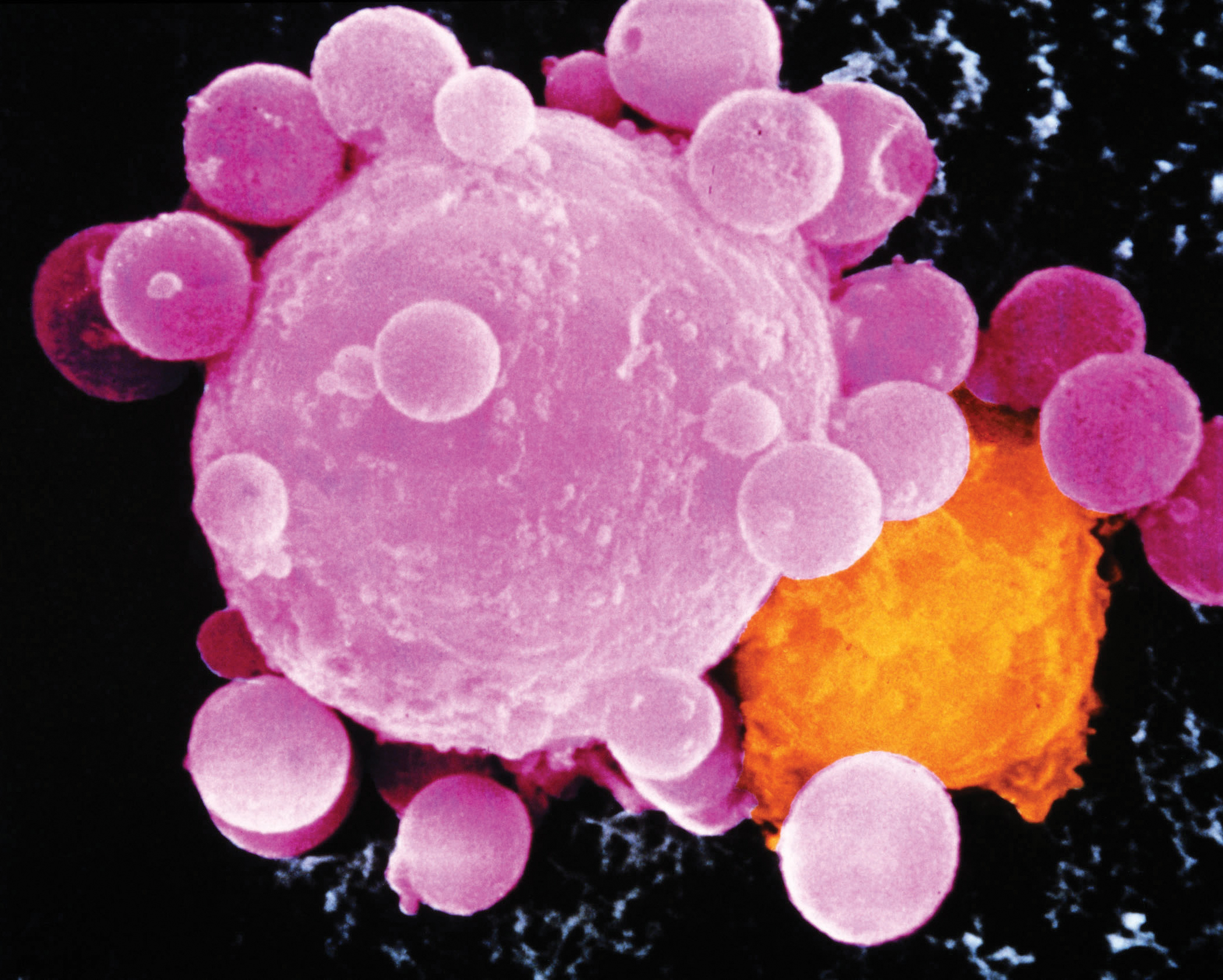
Colored SEM of lymphocyte attacking cancer cell
Caption
Cancer cell death. Colored Scanning Electron Micrograph (SEM) showing a killer T-lymphocyte (orange) inducing a cancer cell (mauve) to undergo Programmed Cell Death (PCD). Mauve vesicles, or apoptotic bodies emerging from the cancer cell indicate PCD (known as apoptosis). Killer T-lymphocytes are part of the body’s immune response system. They are programmed to seek out, attach themselves and kill
Credit
Dr. Andrejs Liepins / Science Photo Library
Description
Cancer cell death. Colored Scanning Electron Micrograph (SEM) showing a killer T-lymphocyte (orange) inducing a cancer cell (mauve) to undergo Programmed Cell Death (PCD). Mauve vesicles, or apoptotic bodies emerging from the cancer cell indicate PCD (known as apoptosis). Killer T-lymphocytes are part of the body’s immune response system. They are programmed to seek out, attach themselves and kill cancer cells, usually using chemicals. In this case the lymphocyte has released a chemical which specifically induces apoptosis in the cancer cell. Apoptosis is a natural method of cell death which is being used as a potential treatment for cancer.
Site_Section
Alt Text
Cancer cell death. Colored SEM of lymphocyte attacking cancer cell.
