You are here
May 21, 2024
Unseen details of human brain structure revealed
At a Glance
- Researchers generated a high-resolution map of all the cells and connections in a single cubic millimeter of the human brain.
- The results reveal previously unseen details of brain structure and provide a resource for further studies.
Fully understanding how the human brain works requires knowing the relationships between the various cells that make up the brain. This entails visualizing the brain’s structure on the scale of nanometers in order to see the connections between neurons.
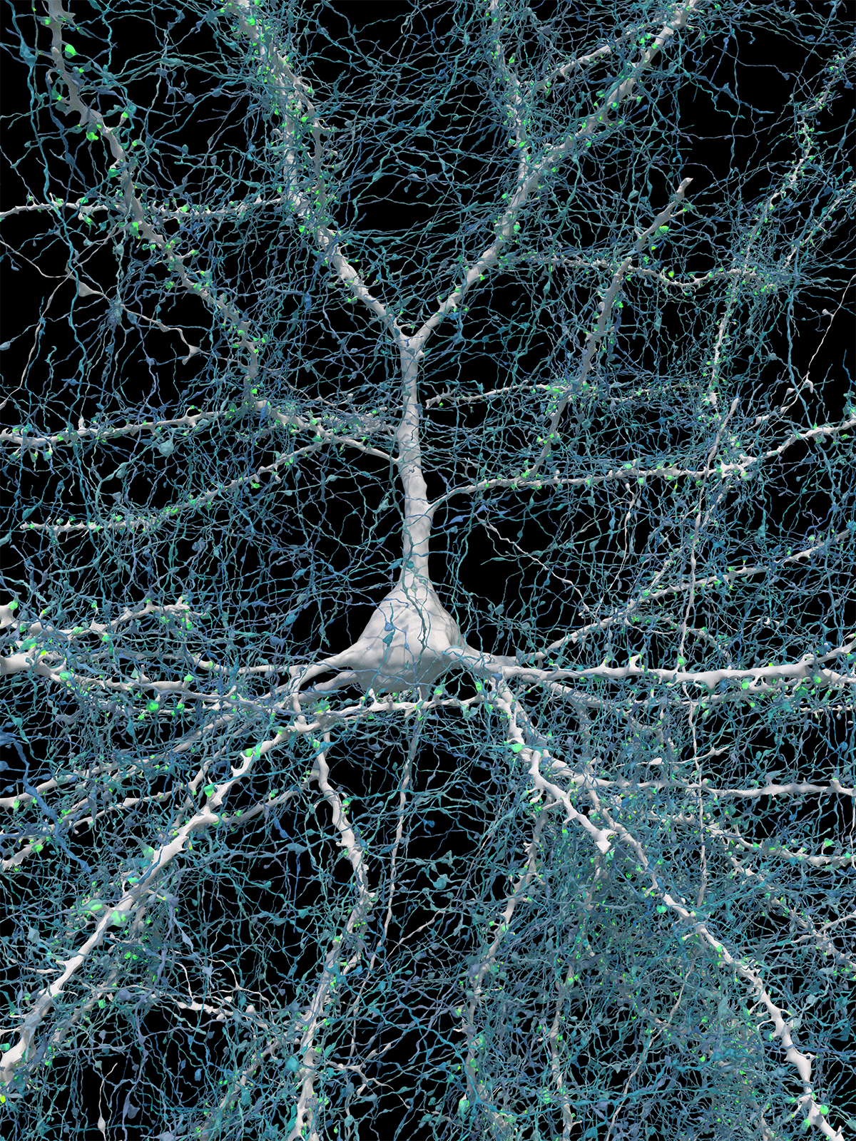
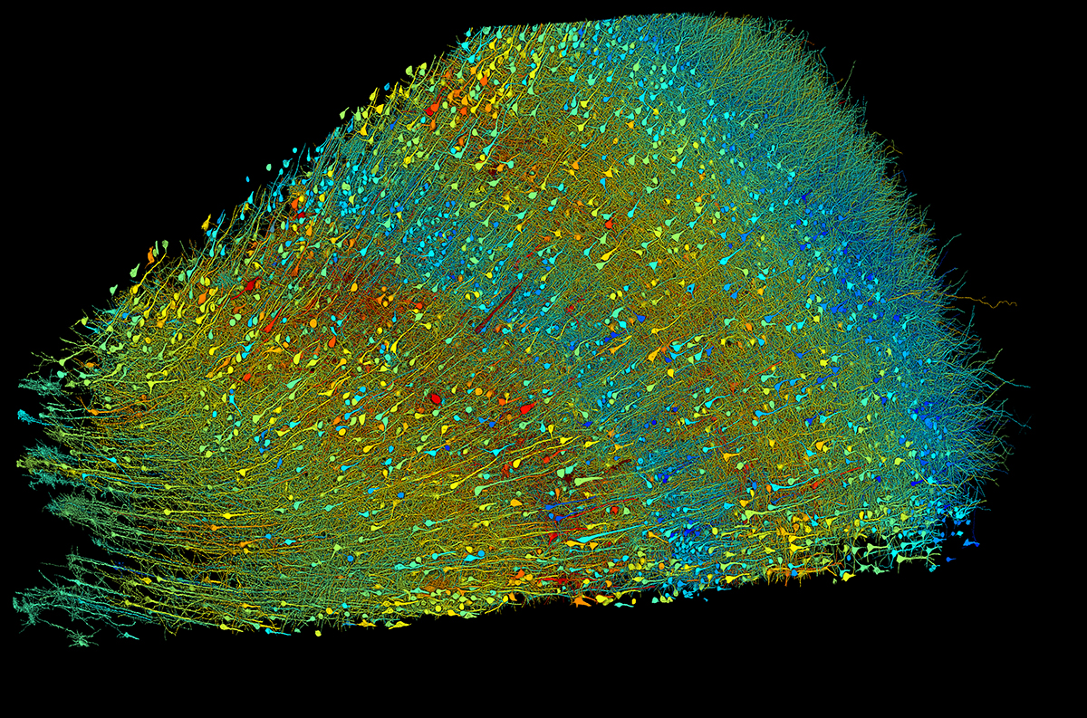
A team of researchers, led by Dr. Jeff Lichtman at Harvard University and Dr. Viren Jain at Google Research, used electron microscopy (EM) to image a cubic millimeter-sized piece of human brain tissue at high resolution. The tissue was removed from the cerebral cortex of a patient as part of a surgery for epilepsy.
The team began by cutting the tissue into more than 5,000 slices, or sections, each of which was then imaged by EM. This yielded about 1.4 petabytes, or 1,400 terabytes, of data. Using these data, the researchers generated a 3D reconstruction of almost every cell in the sample. Results of the NIH-funded study appeared in Science on May 10, 2024.
Analysis of individual cells in the sample revealed a total of more than 57,000 cells. Most of these were either neurons, which send electrical signals, or glia, which provide various support functions to the neurons. Glia outnumbered neurons 2-to-1. The most common glial cells were oligodendrocytes, which provide structural support and electrical insulation to neurons. The one cubic mm sample also contained about 230 mm of blood vessels.
The reconstruction revealed structural details not seen before. The researchers analyzed a type of neuron, called triangular cells, that are found in the deepest layer of the cerebral cortex. Many of these adopted one of two orientations, which were mirror images of each other. The significance of this organization remains unknown.
The team used machine learning to identify synapses—the junctions through which signals pass from one cell to another. They found almost 150 million synapses. Almost all neurons formed only one synapse with a given target cell. But a small fraction formed two or more synapses to the same target. In at least one case, more than 50 synapses connected a single pair of cells. Although rare, connections of seven or more synapses between cells were much more common than expected by chance. This suggests that these strong connections have some functional significance.
The results illustrate just how complex the brain is at the cellular level. They also show the value of connectomics—the science of generating comprehensive maps of connections between brain cells—for understanding brain function.
“The word ‘fragment’ is ironic,” Lichtman says. “A terabyte is, for most people, gigantic, yet a fragment of a human brain—just a miniscule, teeny-weeny little bit of human brain—is still thousands of terabytes.”
The team has made their dataset available to the public. They have also provided various software tools to help examine the brain map. The hope is that further study of the data, by this team and others, will yield new insight into the workings of the human brain.
“This incredible advancement—the ability to capture and process over 1,000 terabytes of data from the brain—wouldn’t have been possible without a study participant’s generous donation and the important partnerships between neuroscientists, computer scientists, and engineers,” says Dr. John Ngai, director of NIH’s BRAIN Initiative. “These collaborations are central in our aim of building a full map of the human brain so we can bring cures closer to the clinic.”
—by Brian Doctrow, Ph.D.
Related Links
- Scientists Build Largest Maps to Date of Cells in Human Brain
- Complete Wiring Map of the Insect Brain
- Mapping the Mammalian Motor Cortex
- Building An Atlas of Brain Function in Mice
- 3-D Model of Human Brain Development and Disease
- An Expanded Map of the Human Brain
- Brain Mapping of Language Impairments
- Brain Basics: Know Your Brain
- The Brain Research Through Advancing Innovative Neurotechnologies® (BRAIN) Initiative
References: A petavoxel fragment of human cerebral cortex reconstructed at nanoscale resolution. Shapson-Coe A, Januszewski M, Berger DR, Pope A, Wu Y, Blakely T, Schalek RL, Li PH, Wang S, Maitin-Shepard J, Karlupia N, Dorkenwald S, Sjostedt E, Leavitt L, Lee D, Troidl J, Collman F, Bailey L, Fitzmaurice A, Kar R, Field B, Wu H, Wagner-Carena J, Aley D, Lau J, Lin Z, Wei D, Pfister H, Peleg A, Jain V, Lichtman JW. Science. 2024 May 10;384(6696):eadk4858. doi: 10.1126/science.adk4858. Epub 2024 May 10. PMID: 38723085.
Funding: NIH’s Brain Research Through Advancing Innovative Neurotechnologies® (BRAIN) Initiative, National Institute of Mental Health (NIMH), National Institute of Neurological Disorders and Stroke (NINDS), and National Institute of Biomedical Imaging and Bioengineering (NIBIB); Stanley Center for Psychiatric Research at the Broad Institute; National Science Foundation; Intelligence Advanced Research Projects Activity.


