You are here
November 19, 2012
Brain Wave Synchronization Key to Working Visual Memory

Short-term memories are stored as synchronized signals between 2 key brain hubs, according to a new study in monkeys. The findings show for the first time how the brain stores visual information for working memory tasks.
Working memory is the ability to hold information in mind and quickly process that information in order to act on it. The visual working memory system has been extensively studied in monkeys and humans. Previous research showed that 2 brain areas, the prefrontal cortex and the parietal cortex, are activated when working memory is used. Electrical signals between neurons in this circuit become synchronized during an identity-matching task in which the subject is asked to match a previously seen object after a time delay.
In the new study, a team led by Dr. Charles Gray at Montana State University analyzed signals from groups of neurons in the prefrontal and parietal brain regions in monkeys performing visual working memory tasks. To earn a juice reward, the monkeys had to correctly identify an object (or its location) that had been previously shown to them on a computer screen. The study was funded by NIH’s National Institute of Mental Health (NIMH) and National Institute of Neurological Disorders and Stroke (NINDS). It appeared online on November 1, 2012, in Science.
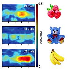
The researchers expected to see increases in synchronous activity, or coherence, between the 2 brain hubs when the object disappeared from the screen and the monkeys had to hold that information in mind. Surprisingly, they found that the degree of coherence depended on the identity of the object. For example, neurons were more in-sync in response to an image of bananas than to an image of a teddy bear. This suggests that these neurons may be selective for particular visual features and that synchronized signals in this circuit carry content-specific information.
“This work demonstrates, for the first time, that there is information about short-term memories reflected in in-sync brainwaves,” says Gray.
Previously, many researchers had thought that the firing rate of single neurons in the prefrontal cortex, the brain’s executive hub, is the major player in working memory. The study found, however, that the parietal cortex was more influential in governing the patterns of synchronization.
The coherence patterns were different for each image that the monkeys saw. This suggests that it may be possible to determine the identity of an object and its location by analyzing these brain waves. Scientists might thus be able to predict the image seen by a monkey, in essence ‘seeing’ their short-term visual memories.
“The Holy Grail of neuroscience has been to understand how and where information is encoded in the brain. This study provides more evidence that large scale electrical oscillations across distant brain regions may carry information for visual memories,” says NIMH Director Dr. Thomas R. Insel.
Related Links
- Mental Replay in Learning and Memory
- Monitoring the Brain’s Memory-Making Cells
- Uncovering the Molecular Basis of Learning and Memory
- Brain Basics: Know Your Brain
References: Science. 2012 Nov 1. [Epub ahead of print]. PMID: 23118014.
