You are here
methods-for-viewing-heart.jpg

Media Folder
Title
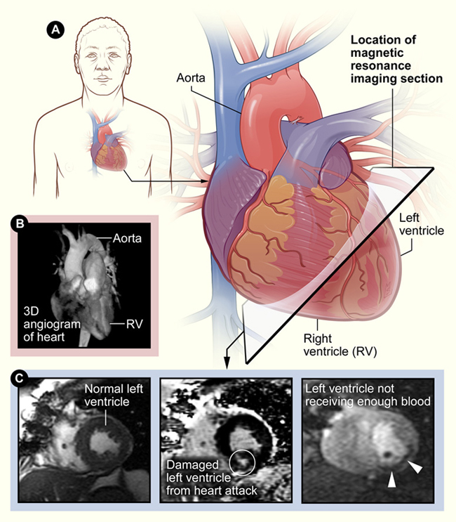
3 panel image showing different methods of viewing the heart
Caption
Panel A: An illustration showing how MRI can take cross-sectional images at any angle through the body. Panel B: A 3-D angiogram examining blood vessels coming off the aorta, the artery that connects the output of the heart to the rest of the body. Panel C: Three MRIs of the heart of a patient with a small heart attack, and who has much more heart muscle at risk.
Credit
NHLBI
Description
Panel A: An illustration showing how MRI can take cross-sectional images at any angle through the body. Panel B: A 3-D angiogram examining blood vessels coming off the aorta, the artery that connects the output of the heart to the rest of the body. Panel C: Three MRIs of the heart of a patient with a small heart attack, and who has much more heart muscle at risk. The left image is a frame from a cine MRI used to make video loops of how well the heart contracts and functions. The middle image is a contrast-enhanced MRI of a heart attack. Living heart muscle appears black while the region damaged by heart attack (inside the circle) appears bright. The right image is a cine MRI frame of the perfusion, or blood supply, of the heart. This shows a large part of the heart is at risk for further damage (dark region marked with arrows).
Site_Section
Alt Text
3 panel image showing different methods of viewing the heart.
