You are here
March 25, 2013
Immune Cells Regulate Brain Development

Scientists discovered a new role for immune cells called microglia in the developing brain. The finding may reveal insights into neurodevelopmental disorders like autism and schizophrenia.
Microglia serve as a primary defense against infection and disease in the central nervous system. In the adult brain, microglia survey the environment, hunting for infectious agents or injured cells. When they detect damage or pathogens, microglia become activated and change shape in order to engulf and eat their targets. They rapidly clear away dying cells and repair tissue damage.
Microglial cells are also found in the brain’s cerebral cortex during prenatal development, but their role wasn’t well understood. To investigate, a research team led by Dr. Stephen Noctor at the University of California, Davis, studied microglia in the developing brain tissue of fetal rats, monkeys and humans. The work, funded in part by NIH’s National Institute of Mental Health (NIMH), appeared in the March 6, 2013, issue of the Journal of Neuroscience.
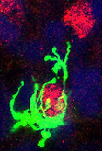
During prenatal development, neural precursor cells — cells with the ability to generate neurons — are rapidly produced in well-defined regions of the brain called proliferative zones. By labeling neural precursor cells and microglia with specific antibodies and other markers, Noctor and his colleagues were able to track them using confocal microscopy.
To their surprise, the scientists found that microglia were engulfing healthy precursor cells in developing brain tissue. Microglia were colonizing proliferative zones and the vast majority of them (over 95%) were activated. Time-lapse images showed microglia contacting healthy neural precursor cells and eating them within a few hours.
The researchers speculated that microglial activity during brain development might affect the number of neural precursor cells. To explore this idea, they used bacterial lipopolysaccharide (LPS) — a toxin that activates microglia — and the antibiotic doxycycline (Dox), which inhibits microglial activation.
The brain tissue of LPS-treated rat pups showed reductions in the number of neural precursor cells of as much as 40% compared to untreated rats. The brain tissue of Dox-treated pups, on the other hand, showed an increase of 20% compared to controls.
Finally, the researchers tested a chemical that selectively kills microglia to see what effect it would have on neural precursor cells. The treatment removed 90% of microglia from the developing brain and significantly increased the neural precursor cell population.
Noctor proposes that the reason microglia feast on neural precursor cells may be to prune brain size during development. “It’s kind of like putting breaks on the system; it’s as if the brain is saying we have enough cells, we don’t need any more and the microglia come in and wipe up the remaining precursor cells,” he says.
Past studies have linked infections and immune activation during pregnancy with neurodevelopmental disorders such as schizophrenia and autism. Future research will explore whether the process this study uncovered could play a role.
— by Meghan Mott, Ph.D.
Related Links
- Prenatal Inflammation Linked to Autism Risk
- How Immune Cells Change Wiring of the Developing Mouse Brain
- Brain Basics
References: J Neurosci. 2013 Mar 6;33(10):4216-33. doi: 10.1523/JNEUROSCI.3441-12.2013. PMID: 23467340.
