You are here
November 8, 2016
Re-envisioning the endoplasmic reticulum
At a Glance
- Advanced imaging techniques revealed a more accurate picture of how the peripheral endoplasmic reticulum is structured.
- The findings may yield new insights for genetic diseases affecting proteins that help shape the endoplasmic reticulum.

Each small structure, or organelle, inside a cell has a specific function. The endoplasmic reticulum (ER) is an organelle that makes and distributes many substances the cell needs, such as proteins, lipids, and sugars. Several proteins help shape the ER’s structure within the cytoplasm (gel-like fluid) of a cell. Mutations in these ER-shaping proteins have been linked to diseases such as hereditary spastic paraplegia—a group of genetic disorders characterized by progressive weakness and stiffness of the legs.
The ER is a complex structure, reaching from the nucleus to the outer edges of the cell. It has many interconnected but structurally distinct domains. One forms a protective membrane surrounding the nucleus. Another, the “peripheral” ER, extends out across the cell. The peripheral ER is conventionally described as an interconnected series of tubule- and sheet-shaped structures. However, traditional microscopy tools have limited how well scientists can visualize the organelle.
To get a closer look, a team led by Dr. Craig Blackstone at NIH’s National Institute of Neurological Disorders and Stroke (NINDS) and Dr. Jennifer Lippincott-Schwartz—formerly at NIH’s Eunice Kennedy Shriver National Institute of Child Health and Human Development (NICHD) and now at the Howard Hughes Medical Institute—used a number of emerging superresolution microscopy techniques to piece together a more detailed picture of the organelle. Results were published in Science on October 28, 2016.
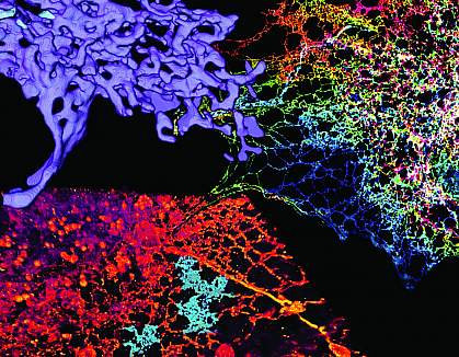
Using techniques based on structured illumination microscopy (SIM), the team tracked the movements of tubules and sheets in the peripheral ER of live cells. SIM is a high-speed technology that allows researchers to rapidly capture molecules in motion. Both regions showed similar, highly dynamic movements. Using these techniques and focused-ion beam scanning electron microscopy, ER sheets didn’t appear as one continuous area. Instead, sheet regions were riddled with spaces that were dynamically changing.
Superresolution imaging across the ER revealed that proteins and lipids are packed into a dense network of tubular-shaped structures―even in areas that have been conventionally classified as sheets. Confocal microscopy revealed that ER-shaping proteins called ATL GTPases, which are characteristic of tubules, are also found scattered throughout sheets.
These findings suggest that previous microscopy techniques lacked the resolution to accurately describe certain regions of the peripheral ER, making very fast-moving tubules appear as a sheet.
“From a neurologic point of view, we work on a class of genetic diseases where the patients have mutations in proteins that control the shape of ER,” Blackstone says. “It’s very important for us to understand what normal is so that we can start to assess what sort of changes these patients have and how it might impact the function of this really critical organelle in the cell.”
―by Tianna Hicklin, Ph.D.
Related Links
- Flavivirus Infection Requires Specific Host Genes
- From Nerve Roots to Plant Roots
- What Is a Cell?
- Hereditary Spastic Paraplegia
References:
Increased spatiotemporal resolution reveals highly dynamic dense tubular matrices in the peripheral ER. Nixon-Abell J, Obara CJ, Weigel AV, Li D, Legant WR, Xu CS, Pasolli HA, Harvey K, Hess HF, Betzig E, Blackstone C, Lippincott-Schwartz J. Science. 2016 Oct 28;354(6311). pii: aaf3928. PMID: 27789813.
Funding:
NIH’s Eunice Kennedy Shriver National Institute of Child Health and Human Development (NICHD), National Institute of Neurological Disorders and Stroke (NINDS), National Institute of General Medical Sciences (NIGMS), and Office of AIDS Research; National Key Research Program of China; Howard Hughes Medical Institute; and University College London School of Pharmacy.
