You are here
April 12, 2016
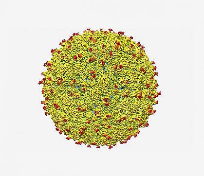
Zika virus structure revealed
At a Glance
- The Zika virus surface is similar to that of dengue and related viruses at the near-atomic level, researchers found, but with a notable difference.
- The structure provides clues to understanding how Zika virus enters human cells and suggests ways to design drugs or vaccines to combat the virus.

Zika virus was discovered in Uganda in 1947. It’s a type of virus called a flavivirus. Other flaviviruses include dengue, yellow fever, and West Nile viruses. Like its relatives, Zika virus is mainly transmitted to humans through the bite of infected Aedes aegypti mosquitoes. Most people infected with Zika virus don’t get sick. The 20% or so who do tend to have mild symptoms that subside within a week. Symptoms can include fever, rash, joint pain, and conjunctivitis (pink eye).
Zika virus circulated relatively unnoticed in areas of Africa and Southeast Asia until 2007, when it caused an outbreak in a new region, Yap Island in Micronesia. In 2013, the virus caused an outbreak in French Polynesia. The first confirmed case of infection in Brazil came in May 2015. Since that time, the virus has spread to other countries and territories in Central and South America as well as the Caribbean, including Puerto Rico and the U.S. Virgin Islands. The current outbreak provides mounting evidence that Zika virus can also cause a serious birth defect called microcephaly in babies born to mothers infected with the virus during pregnancy. Microcephaly is a condition marked by an unusually small head, brain damage, and developmental delays.
To learn more about Zika virus, a team led by Drs. Richard Kuhn and Michael Rossmann at Purdue University examined the structure of a mature Zika virus particle at near-atomic resolution. They used a technique called cryo-electron microscopy. The process involves freezing virus particles and firing a stream of high-energy electrons through the sample to create tens of thousands of images. These 2-D images are then combined to yield a detailed 3-D view of the virus. The work was funded by NIH’s National Institute of Allergy and Infectious Diseases (NIAID). The research team also included NIAID investigator Dr. Theodore Pierson. Results appeared online on March 31, 2016, in Science.
The surface of the flavivirus is composed of a shell made of 180 copies of both an envelope glycoprotein and 1 of 2 other proteins anchored in a lipid membrane. The researchers found that the Zika virus structure is similar to that of other known flaviviruses, except for one region of the envelope glycoprotein. Flaviviruses may use this glycoprotein region to attach to and enter human cells.
The newly identified Zika variation could help explain the virus’s ability to attack nerve cells. It might also shed light on Zika’s proposed link to microcephaly. If the site on the envelope glycoprotein functions in Zika as it does in related viruses, the detailed structure might help scientists design ways to block viral attachment and entry to human cells.
“The structure of the virus provides a map that shows potential regions of the virus that could be targeted by a therapeutic treatment, used to create an effective vaccine or to improve our ability to diagnose and distinguish Zika infection from that of other related viruses,” Kuhn says.
Related Links
- Zika Virus
- Zika Virus Information and Resources
- Video: Zika Virus: A Pandemic in Progress
- Block the Buzzing, Bites, and Bumps: Preventing Mosquito-Borne Illnesses
- Microcephaly Information Page
References: The 3.8 Å resolution cryo-EM structure of Zika virus. Sirohi D, Chen Z, Sun L, Klose T, Pierson TC, Rossmann MG, Kuhn RJ. Science. 2016 Mar 31. pii: aaf5316. [Epub ahead of print]. PMID: 27033547.
Funding: NIH’s National Institute of Allergy and Infectious Diseases (NIAID).
