You are here
March 14, 2017
3-D printer creates blood vessel networks
At a Glance
- A research team developed a new 3-D printing system for fabricating blood vessel networks.
- With further development, the system could be used to engineer tissues and their associated blood vessel networks.

Networks of blood vessels branch throughout almost every tissue of the body. These vessels are channels for the transport of blood cells, nutrients, and waste products. Researchers have made much recent progress in engineering new tissues and organs. However, creating functional networks of blood vessels to provide vital transport to the cells within these engineered tissues remains a major challenge.
Engineers have been using 3-D printers in the lab to try to make models of, and even replacements for, many parts of the body, including blood vessels. 3-D printers can assemble raw materials into very complex products. Researchers had previously fabricated a single blood vessel, which amounted to no more than a long and slender tube. The next hurdle is to create complex, branching networks of blood vessels.
A team of engineers led by Dr. Shaochen Chen of the University of California, San Diego, aimed to improve on current 3-D printers to better engineer complex tissues like blood vessel networks. Their research was supported by NIH’s National Institute of Biomedical Imaging and Bioengineering (NIBIB). Results were published online in advance of the April 2017 issue of Biomaterials.
Chen’s team devised a system that used digital 3-D designs from a computer to guide a 3-D printer’s fabrication of blood vessels using biocompatible hydrogel materials. The device relied on ultraviolet light and about 2 million microscopic mirrors to guide the polymerization of the hydrogel materials into particular solid shapes. The designs from the computer dictated the angles of the microscopic mirrors to print the 3-D shape layer by layer.
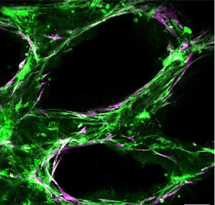
The engineers created 2 versions of their 3-D blood vessel network. One was made using 2 hydrogels. The other was made with the 2 hydrogels plus 2 types of living cells. Each version measured about 4 mm by 5 mm (about the size of a pencil eraser) and was only 0.6 mm thick.
The team implanted both versions under the skin of mice. Within 2 weeks, the engineered blood vessel network made with the living cells had joined with the mouse blood vessel network, allowing blood to circulate through. Less evidence of this joining was seen in the version made without living cells.
“Almost all tissues and organs need blood vessels to survive and work properly. This is a big bottleneck in making organ transplants, which are in high demand but in short supply,” says Chen. “3-D bioprinting organs can help bridge this gap, and our lab has taken a big step toward that goal.”
The results show that a complex tissue resembling blood vessels can be formed using a 3-D printer. The ultimate challenge for this research team is to engineer heart tissue with a complex network of blood vessels. Such tissues might be used to replace damaged heart muscle or for drug testing.
—by Geri Piazza
Related Links
- Bioengineered Blood Vessel Grafts Grow in Animals
- Repairing Nerve Pathways With 3-D Printing
- Stem Cells Coaxed To Create Working Blood Vessels
- Fixing Flawed Body Parts: Engineering New Tissues and Organs
- Tissue Engineering and Regenerative Medicine
- Circulation and Blood Vessels
References: Direct 3D bioprinting of prevascularized tissue constructs with complex microarchitecture. Zhu W, Qu X, Zhu J, Ma X, Patel S, Liu J, Wang P, Lai CS, Gou M, Xu Y, Zhang K, Chen S. Biomaterials. 2017 Apr;124:106-115. doi: 10.1016/j.biomaterials.2017.01.042. PMID: 28192772.
Funding: NIH’s National Institute of Biomedical Imaging and Bioengineering (NIBIB), California Institute for Regenerative Medicine, and National Science Foundation.
